
Precio y stock a confirmar
Ed. Benjamin Cummings, año 2001. Tamaño 28 x 21,5 cm. Incluye más de 400 figuras en blanco y negro. Estado: Usado excelente. Cantidad de páginas: 200
 You may ask why we are creating a third edition. Does anatomy change between editions? With respect to what is taught to students of anatomy, formally or informally, there is not much change. Occasionally, a new variation is noted, but substantively and as a practical matter, anatomy doesn’t change. On a grander scale, the anatomic arrangement of our bodies IS subject to infinitesimal change in an evolutionary sense, but that’s not why we composed a new edition. We did it because it was time to freshen the illustrations and text, to go over material and find new and better ways to illustrate and express the anatomy and its function. We also cleaned up errors and made the presentation more clear.
We worked to improve the visual appeal of the plates, struggling to resist putting 10 lbs. of information in a 5-lb. plate. We found our earlier coverage of joints to be inadequate. We produced nine new plates, five of which make up that deficit: the temporomandibular, shoulder, elbow, sacroiliac, hip, and knee joint plates. We rearranged the order of presentation of material to make it easier for anatomy teachers to integrate our material with commonly employed anatomy texts.
Our visually-based quizzes on bones, arteries, and veins have been expanded to include joints. We reorganized the lists of vessels in those quizzes to make them more digestible. We revised and gave new life to a full third of the existing plates, including expansion of the integument to two plates, updating the plate on HIV-induced immunosuppression, and vastly improving the plates on distribution of spinal nerves, the meninges, the visual system, and the renal tubules. The literature on innervation of skeletal muscle has been reviewed and updated, as has Appendix B.
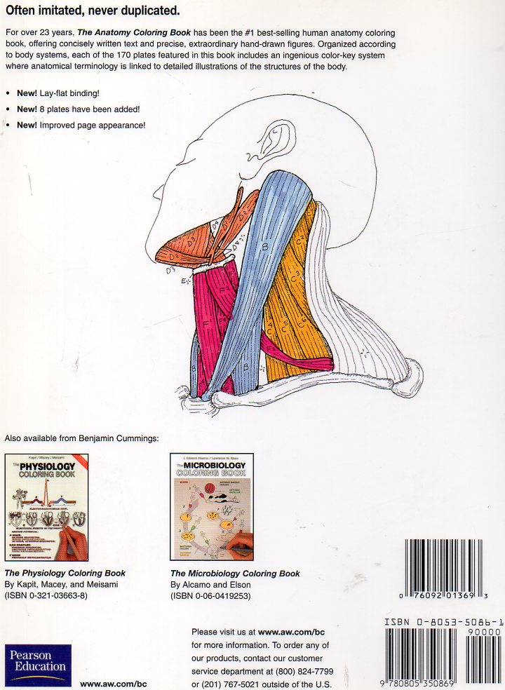
 To borrow from the Preface of the second edition, we are grateful to the thousands of colorers who have advised and encouraged us, including coaches, trainers, teachers, paramedics, body workers, court reporters, attorneys, insurance claims adjusters, judges, and students and practi¬tioners of dentistry and dental hygiene, nursing, medicine/surgery, chiropractic, podiatry, massage therapy, myotherapy, physical therapy, occupational therapy, and exercise therapy. More informal seekers of self-realization and those with impairments have been drawn to The Anatomy Coloring Book because of its lighter, more visual approach. Truly, a picture is worth a thousand words!
HOW THE BOOK IS ARRANGED
The book is divided by subject matter into sections. The sections contain groups of plates, each dealing with a separate topic within that subject heading.
A plate consists of an illustration with various parts to be colored, related titles (also to be colored), an explanatory paragraph of text, and coloring notes (CN).
You can begin with any section, but it is best to color that section in the order In which the plates are presented. Feel free to skip the plates that might be too complex or irrelevant to your area of interest.
 HOW MANY COLORS NEEDED
It is best to have at least 10 pens or pencils (no crayons). Pencils are more versatile because one can lighten or darken each color. Felt-tipped pens, on the other hand, produce brighter colors.
The more colors you have available, the greater the pleasure. If you are able to purchase your colors Individually (as opposed to a set), you should choose mostly light colors, but be sure to Include gray and black.
HOW THE COLORING SYSTEM WORKS
The parts of an illustration that are meant to be colored are drawn with, or separated from each other by, dark outlines. They are also identified by small letter labels (A, B, etc.). The «titles» (names or terms referring to those parts) are printed with outlined letters, followed by the same letter labels. Color a part and its respective title with the same color. Do not use that color again on a different part and title on that plate, unless you run out of colors, and have to repeat some colors.
When different parts of an illustration are related to each other in some fundamental way, they will receive the same letter labels, but with different superscripts (A1, A2) for identification purposes. All those parts will receive the same color.
Occasionally, you will come across a title or general heading that is meant to be colored but does not refer to any specific part of the illustration. In such cases, the small letter label will be followed by a dash (A-, B-), and only the title or heading will be colored.
Areas or words meant to be col ¡red gray are identified by an asterisk (* ); if colored black, by a black circle ( • ); and when not to be colored at all, by the «don’t color» sign
 HOW TO APPROACH EACH PLATE
Whether you read the explanatory material before coloring or vice versa, you should always read the coloring notes (CN) before starting to color. The notes (located at the top of the plate) contain rec-ommendations about which colors to use and what to take notice of when coloring that particular plate.
Begin by coloring the first title in the list of titles to be colored. The title will be followed by a small letter label (A). Locate and color the part of the illustration to which the title refers. It is important that you color the titles in the order that they are presented; they are usually listed that way for specific reasons.
The titles are usually placed away from the illus¬trations to facilitate your review. Try covering them when testing your recall of the material.
It is recommended that you reserve your lightest colors for the largest areas to be colored. A dark color on an especially large part of the illustration would dominate the plate. Certain colors are tradi¬tionally associated with certain structures of the body: red for arteries, blue for veins, purple for capillaries, yellow for nerves, and green for lym¬phatics. Where you are asked to identify a diverse group of such structures (i.e., many different arter¬ies or many veins), you will naturally have to use more than just the one representative color.
TABLE OF CONTENTS
Preface
Acknowledgements
Introduction to coloring (Important tips on how to get the most from this book)
I- ORIENTATION TO THE BODY
1- Anatomic Planes and Sections
2- Terms of Position and Direction
3- Systems of the Body (1)
4- Systems of the Body (2)
5- Regions of the Body: Anterior View
6- Regions of the Body: Posterior View
7- Cavities and Linings
II- CELLS AND TISSUES
8- The Generalized Cell
9- Cell Division/Mitosis
10- Tissues: Epithelium
11- Tissues: Fibrous Connective Tissue
12- Tissues: Supporting Connective Tissue
13- Tissues: Muscle
14- Tissues: Skeletal Muscle Microstructure
15- Tissues: Nervous
16- Neuromuscular Integration
17- Integration of Tissues
III- INTEGUMENTARY SYSTEM
18- The Integument: Epidermis
19- The Integument: Dermis
IV- SKELETAL AND ARTICULAR SYSTEMS
20- Long Bone Structure
21- Axial/Appendicular Skeleton
22- Classification of Joints
23- Terms of Movement
24- Bones of the Skull (1)
25- Bones of the Skull (2)
26- Temporomandibular Joint
27- Vertebral Column
28- Cervical and Thoracic Vertebrae
29- Lumbar, Sacral, Coccygeal Vertebrae
30- Bony Thorax
31- Upper Limb: Pectoral Girdle/Arm Bone
32- Upper Limb: Glenohumeral (Shoulder) Joint
33- Upper Limb: Forearm Bones
34- Upper Limb: Elbow Joints
35- Upper Limb: Wrist and Hand Bones & Joints
36- Upper Limb: Bones/Joints in Review
37- Lower Limb: Hip Bone, Pelvic Girdle and Pelvis
38- Lower Limb: Male and Female Pelves
39- Lower Limb: Sacroiliac and Hip Joints
40- Lower Limb: Thigh and Leg Bones
41- Lower Limb: Knee Joint
42- Lower Limb: Ankle and Foot Bones
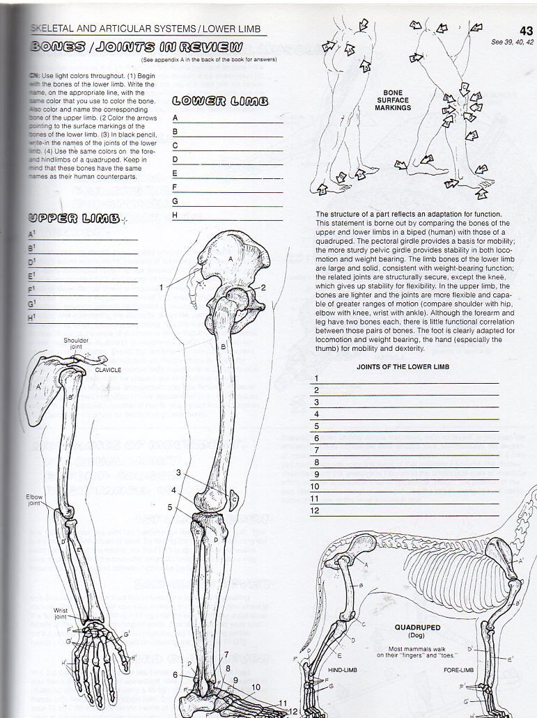
43- Lower Limb: Bones/Joints in Review
V- SKELETAL MUSCULAR SYSTEM
44- Introduction to Skeletal Muscle
45- Integration of Muscle Action
46- Head: Muscles of Facial Expression
47- Head: Muscles of Mastication
48- Neck: Anterior and Lateral Muscles
49- Torso: Deep Muscles of Back and Posterior Neck
50- Torso: Muscles of Thorax and Posterior Abdominal Wall
51- Torso: Muscles of Anterior Abdominal Wall and Inguinal Regii
52- Torso: Muscles of the Pelvis
53- Torso: Muscles of the Perineum
54- Upper Limb: Muscles of Scapular Stabilization
55- Upper Limb: Musculotendinous Cuff
56- Upper Limb: Movers of Shoulder Joint
57- Upper Limb: Movers of Elbow and Radioulnar Joints
58- Upper Limb: Movers of Wrist & Hand Joints
59- Upper Limb: Movers of Hand Joints (Intrinsics)
60- Muscles of the Upper Limb in Review
61- Lower Limb: Muscles of the Gluteal Region
62- Lower Limb: Muscles of the Posterior Thigh
63- Lower Limb: Muscles of the Medial Thigh
64- Lower Limb: Muscles of the Anterior Thigh
65- Lower Limb: Muscles of the Anterior & Lateral Leg
66- Lower Limb: Muscles of the Posterior Leg
67- Lower Limb: Muscles of the Foot (Intrinsics)
68- Muscles of the Lower Limb in Review
69- Functional Overview
VI- NERVOUS SYSTEM
70- Organization
71- Functional Classification of Neurons
72- Synapses and Neurotransmitters
VII- CENTRAL NERVOUS SYSTEM
73- Cerebral Hemispheres
74- Tracts and Nuclei of Cerebral Hemispheres
75- Diencephalon
76- Brain Stem/Cerebellum
77- Spinal Cord
78- Ascending Tracts
79- Descending Tracts
VIII- CNS: CAVITIES & COVERINGS
80- Ventricles of the Brain
81- Meninges
82- Circulation of Cerebrospinal Fluid (CSF)
IX- PERIPHERAL NERVOUS SYSTEM
83- Cranial Nerves
84- Spinal Nerves and Nerve Roots
85- Spinal Reflexes
86- Distribution of Spinal Nerves and Thoracic Spinal Nerve
87- Cervical Plexus and Nerves to the Neck
88- Brachial Plexus and Nerves to the Upper Limb
89- Lumbosacral Plexus and Nerves to the Lower Limb
90- Dermatomes
91- Sensory Receptors
X- AUTONOMIC OR VISCERAL NERVOUS SYSTEM
92- ANS: Sympathetic Division (1)
93- ANS: Sympathetic Division (2)
94- ANS: Parasympathetic Division
XI- SPECIAL SENSES
95- Visual System (1).
96- Visual System (2)
97- Visual System (3)
98- Auditory apd Vestibular Systems (1)
99- Auditory and Vestibular Systems (2)
100- Taste and Olfaction
XII- CARDIOVASCULAR SYSTEM
101- Blood and Blood Elements
102- Scheme of Blood Circulation
103- Bloodvessels
104- Mediastinum, Walls, and Covering of the Heart
105- Chambers of the Heart; Circulation Through the Heart
106- Cardiac Conduction System and the ECG
107- Coronary Arteries and Cardiac Veins
108- Arteries of the Head and Neck
109- Arteries of the Brain
110- Arteries and Veins of the Upper Limb
111- Arteries of the Lower Limb
112- Aorta and Branches
113- Arteries to Gastrointestinal Tract and Related Organs
114- Arteries of the Pelvis and Perineum
115- Review of Principal Arteries
116- Veins of the Head and Neck
117- Caval and Azygos Systems
118- Veins of the Lower Limb
119- Hepatic Portal System
120- Review of Principal Veins
XIII- LYMPHATIC SYSTEM
121- Lymphocyte Circulation
XIV- IMMUNE (LYMPHOID SYSTEM)
122- Introduction
123- Natural and Acquired Immunity
124- Thymus and Red Marrow
125- Spleen
126- Lymph Node
127- Mucosal Associated Lymphoid Tissue (M.A.L.T.)
128- HIV-lnduced Immunosuppression
XV- RESPIRATORY SYSTEM
129- Overview of the System
130- External Nose, Nasal Septum, and Nasal Cavity
131- Paranasal Air Sinuses
132- Pharynx and Larynx
133- Lobes and Pleurae of the Lungs
134- Lower Respiratory Tract
135- Mechanism of Respiration
XVI- DIGESTIVE SYSTEM
136- Overview of the System
137- Oral Cavity and Relations
138- Anatomy of a Tooth: Adult/Child Dentition
139- Pharynx and Swallowing
140- Peritoneum
141- Esophagus and Stomach
142- Small Intestine *
143- Large Intestine
144- Liver
145- Biliary System and Pancreas
XVII- URINARY SYSTEM
146- Urinary Tract
147- Kidneys and Related Retroperitoneal Structures
148- Kidney and Ureter
149- Renal Tubule
150- Tubular Function and Renal Circulation
XVIII- ENDOCRINE SYSTEM
151- Introduction
152- Pituitary Gland and Hypothalamus
153- Pituitary Gland and Target Organs
154- Thyroid and Parathyroid Glands
155- Adrenal (Suprarenal) Glands
156- Pancreatic Islets
XIX- REPRODUCTIVE SYSTEM
157- Male Reproductive System
158- Testis
159- Male Urogenital Structures
160- Female Reproductive System
161- Ovary
162- Uterus, Uterine Tubes, and Vagina
163- Menstrual Cycle
164- Breast (Mammary Gland)
XX- HUMAN DEVELOPMENT
165- Development of Embryo (1)
166- Development of Embryo (2)
167- Embryo/Fetus Coverings
168- Endochondral Ossification
169- Development of Central Nervous System
170- Fetal Circulation
BIBLIOGRAPHY AND REFERENCES
APPENDIX A: ANSWER KEYS (PLATES 36, 43, 60, 68, 115, 120)
APPENDIX B: INNERVATION OF SKELETAL MUSCLES
GLOSSARY
INDEX